
Ever wondered why some wounds on the equine lower limb take so long to heal?? Read about the process of wound healing in following article written by Dr Erica Schmidt about her delightful patient “Dolly”!
“Dolly” is a yearling Quarter Horse filly that lacerated her right hind leg in a fence. The laceration extended from the lower portion of the hock joint to roughly halfway down the cannon bone on the front side of the leg. Luckily, the wound appeared to be isolated to the skin and superficial connective tissue without extending into the joint, which would have created a much more serious problem. At the initial vet visit shortly after the wound occurred, the skin that had covered the area was mostly gone, with just a flap of skin attached the bottom portion of the wound. Unfortunately, the blood supply to the area was compromised and the skin was likely to die, so it was surgically removed. There was not enough remaining skin to suture the wound closed, so it was cleaned and bandaged.
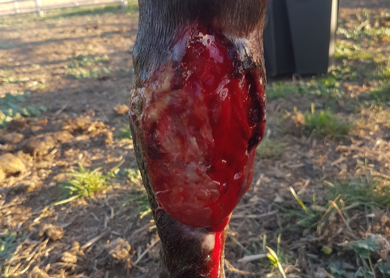
The initial phase of wound healing is known as acute inflammation. This phase occurs during the first three days after the wound occurs, during which, the body is focused on stopping bleeding, preventing infection, and establishing a healthy population of inflammatory cells in the area. During the acute inflammation stage of Dolly’s wound, it had been cleaned and bandaged daily to protect the wound and prevent infection. Dolly was also placed on antibiotics to further prevent infection in the area, as well as an anti-inflammatory to relieve pain and excessive swelling.
Acute inflammation in Dolly’s wound. New granulation tissue is visible in the lower left side.
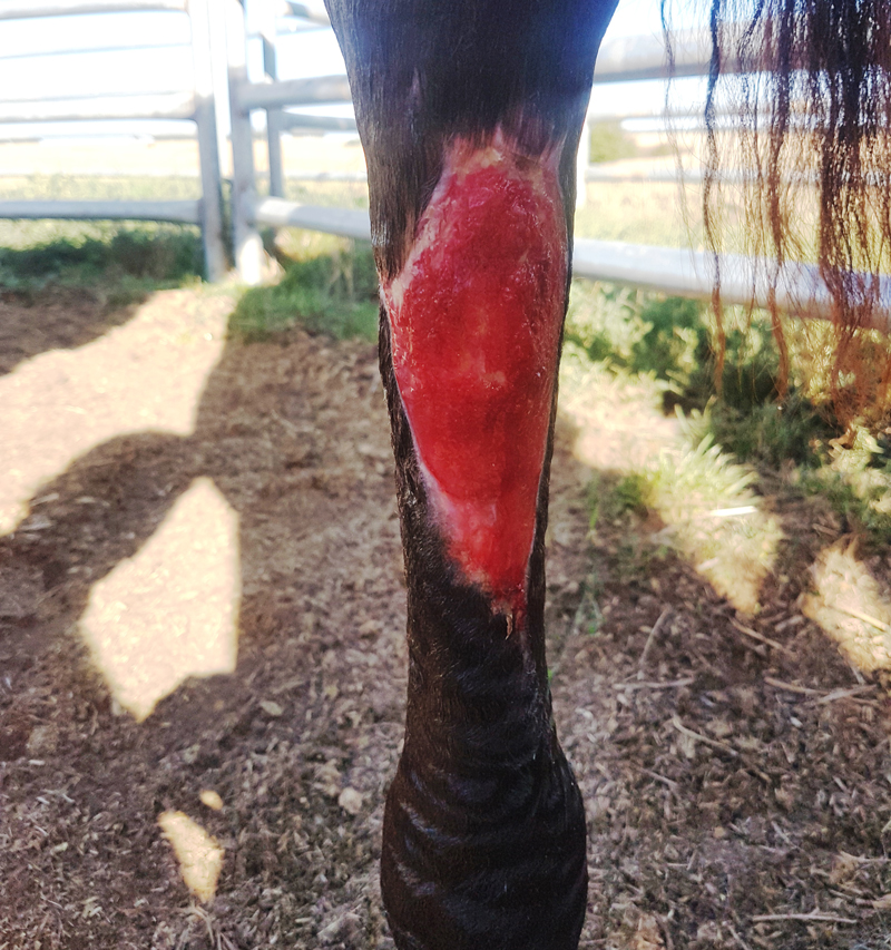
Healthy granulation tissue surrounded by new epithelium (skin).
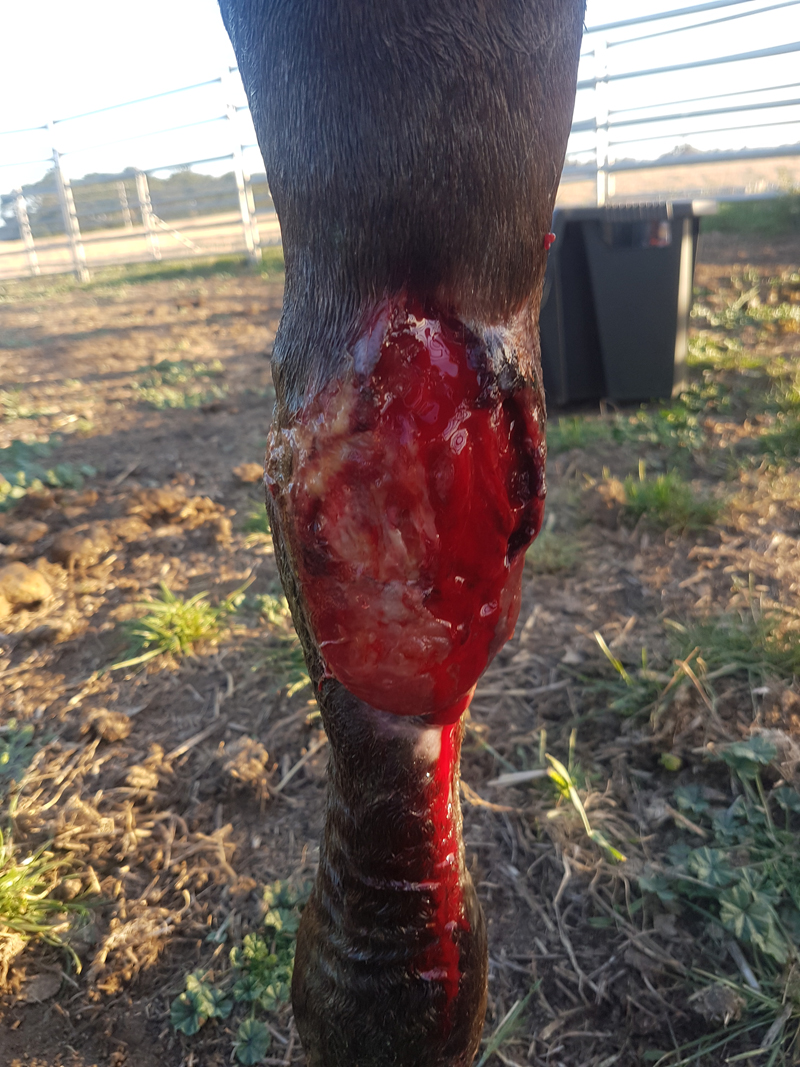
The second phase of wound healing, which takes place over days 3-14 after the wound occurs, is known as the cellular proliferation phase. Granulation tissue is formed during this time, which provides a foundation for new blood vessels, nerves, and skin to be produced. As many horse owners know, granulation tissue must be managed carefully in order to avoid excessive production, known as proud flesh, which can actually delay healing. Dolly’s bandage was changed every 2-3 days during the proliferation phase. During each bandage change, the wound was cleaned and analysed. The discharge from the wound remained clear to bloody in colour, confirming that no infection was present. The granulation tissue was maintained at a healthy level using a strategic combination of steroid wound cream and bandage pressure.
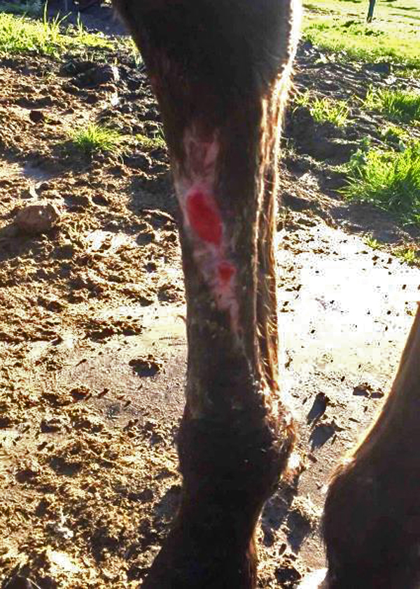
The third and final phase of wound healing is known as remodelling. This phase overlaps with the proliferative phase, beginning at approximately 7 days post-injury. During remodelling, the bed of granulation tissue over the wound becomes infiltrated with new blood vessels, nerves, and connective tissues. Skin re-growth also begins during this phase, with new epithelial cells lining the outside of the wound. Once remodelling was evident in Dolly’s wound, bandage changes decreased in frequency to twice weekly. Since steroids (which were used to supress excess granulation tissue) are known to slow down the remodelling process, the steroid wound cream was alternated with another anti-inflammatory wound dressing that didn’t contain a steroid. The bandages were able to be taken off completely when the entire wound was covered in new epithelium and no more granulation tissue remained visible. Since then, most of the hair has grown back over the scar and Dolly is doing great!
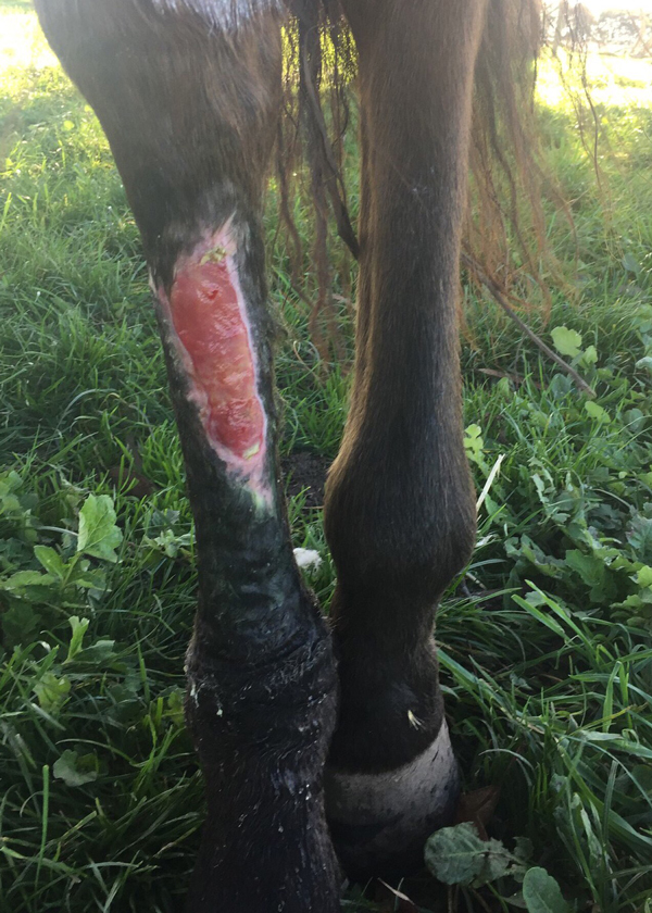
Continued remodelling of wound.
Nearly done!